Xr L Spine Complete
Posted By admin On 24/07/22Xr bone survey complete 77075 xr calcaneus: 73650 xr chest 1 view 71010: xr chest 2 views 71020: xr chest 2vw w/apical lordotic 71021 xr chest 2vw w/obliques: 71022 xr chest 4+vw min 71030: xr chest spec view obl/lor/dec 71035: xr clavicle 73000 xr c-spine 1 vw: 72020 xr c-spine ap&lat 2-3 view std 72040: xr c-spine comp obl/flex/ext 72052: xr. The most commonly ordered spine radiographs, x-rays of the cervical spine are used to evaluate trauma and everyday neck pain. X-rays are also useful for evaluation of the postoperative patient. The three essential views are AP, Lateral, and Odontoid. The AP view of the cervical spine is shown here without comment. 72052 Cervical Spine, Complete 72070 Thoracic Spine (2 views) 72072 Thoracic Spine (3 views) 72100 Lumbar Spine (2 or 3 views) 72110 Lumbar Spine, Complete 72114 Lumbar, Complete (w/ Flex & Ext). L-spine complete with bending, L spine w/ flex/ext, L-spine complete w/ flex/ext 72069 CR Scoliosis 2-4 Scoliosis screening, Scoli, Thoraco-lumbar for scoliosis 72220 CR Sacrum Coccyx Min 2V 2-3 Sacrum/Coccyx, sacral spine.
Clues to lumbar pathology are found in all views: AP, Lateral, Obliques, and Flexion/extension views.
The AP view
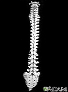
The AP view may reveal scoliosis or spine mets. The scoliosis of adolescence is usually rightward in the thoracic spine (away from the heart), and leftward in the compensatory lumbar curve. The lumbar scoliosis of the elderly, degenerative spine can be in either direction. Keep in mind that the convexity *opens* the neural foramen, and the concavity *narrows* the foramen. The concave part of the scoliosis can close the neural foramen and pinch the nerve. While looking at the AP, be sure to visualize the pedicles at every level. A “winking owl” sign may herald a destructive process like metastatic cancer in the pedicle.
The Lateral view
The lateral view may show fracture or subluxation. Each vertebra should be examined in the lateral view, looking for a compression fracture, often seen in the upper lumbar or lower thoracic, sometimes representing an old or occult fracture. Next look at the posterior vertebral (marginal) line, looking for the subluxation of spondylolisthesis, most often occurring at L4-5, but possible at any level.
The x-rays, of course, do not image the discs, but you can make inferences about the disc based on the space between the vertebrae. L4-5 should be the tallest disc, with L3-4 and L5-S1 tied for second place. Loss of disc height is radiographic evidence of disc degeneration.

The oblique view
The oblique view is useful for one finding only: Pars fracture (defect). If there is a defect of the pars interarticularis, it may be imaged on the oblique view, and will appear as a Scottie Dog sign. At each vertebra, look for the Scottie Dog. If the Scottie Dog has a “broken neck,” you are looking at a fractured pars interarticularis.
Flexion/extension views

If a spondylolisthesis is present, you may see anterior displacement of one vertebra upon another. In an unstable, mobile, spondylolisthesis, the “slip” will be greater in flexion, and less on extension. In other words, the “slip” may reduce on extension. In any case, a mobile spondylolistheis can cause severe mechanical back pain, with leg pain if a nerve is pinched in the intervertebral foramen.
Order plain lumbar radiographs to evaluate back pain and trauma. They yield great information about the vertebrae and spinal configuration.
The lumbar spine flexion and extension views images the lumbar spine which consists of five vertebrae.
Indications
These views are specialized projections to provide functional tests 1 of lumbar spine instability, often in the context of spondylolisthesis.
Patient position
- the patient is positioned erect:
- ideally, spinal imaging should be taken erect in the setting of non-trauma to give a functional overview of the lumbar spine
- all imaging of patients with suspected spinal injury must occur in the supine position without moving the patient
- in the lateral decubitus position, position the patient so that the humeri are extended 90 degrees to the thorax, with the elbows flexed so that the forearms are parallel to the thorax. Spinal curvature in the AP projection will determine if a right lateral or a left lateral is performed.
- when implementing horizontal beam technique, ensure the distal upper limbs are not overlying the region of interest. Ask the patient to cross their arms over their upper thorax, or to extend them in a similar position to that achieved in the lateral decubitus position
- flexion
- at the last possible moment, instruct the patient to 'bend forward' from the lower back, flexing their lower spine
- extension
- at the last possible moment, instruct the patient to 'lean back' from the lower back essentially extending their lower spine
Technical factors
- lateral projection
- expiration (to minimize superimposition of the diaphragm over the upper lumbar spine)
- centering point
- the level of the iliac crest
- coronal centering point is directly over the lumbar vertebra, which corresponds to the posterior third of the abdomen
- the central ray is perpendicular to the image receptor
- collimation
- superiorly to include T12/L1
- inferior to include the sacrum
- anterior to include the anterior border of the lumbar vertebral bodies
- posterior to include all elements of the posterior column, particularly the spinous processes
- orientation
- portrait
- detector size
- 35 cm x 43 cm
- exposure
- 70-80 kVp
- 60-80 mAs
- SID
- 110 cm
- grid
- yes (ensure the correct grid is selected if using focused grids)
Image technical evaluation
Xr L Spine Complete Series Views Cpt Code
- annotations affixed to demonstrate flexion and extension
- the entire lumbar spine should be visible from T12/L1 to L5/S1
- adequate image penetration and image contrast is evident by clear visualization of lumbar vertebral bodies, with both trabecular and cortical bone demonstrated
Practical points
- physical demonstration of the projection is often best to ensure patient fully understands the procedure
- ensure centering is adjusted when the patient moves into position
- utilize an erect bucky when performing horizontal beam laterals to utilize oscillating grids, automatic expose control, and CR/IR alignment
- if the patient demonstrates spinal scoliosis, ensure that the side with the convexity is closest to the IR. This will utilize the diverging beam and aid in achieving superimposition of the upper and lower endplates
- try to remove as many possible image artefacts, especially when performing horizontal beam technique in a trauma context
- if using a CR system, a smaller cassette 30 x 35 can be used when the sacral region does not need to be demonstrated. When centering, place the height of the CR 2.5 cm above the iliac crests
- 1. Boyle Cheng, Anthony E. Castellvi, Reginald J. Davis, David C. Lee, Morgan P. Lorio, Richard E. Prostko, Chip Wade. Variability in Flexion Extension Radiographs of the Lumbar Spine: A Comparison of Uncontrolled and Controlled Bending. (2016) International Journal of Spine Surgery. 10: 20. doi:10.14444/3020 - Pubmed
- 2. Justin R Câmara, Joseph R. Keen, Farbod Asgarzadie. Functional Radiography in Examination of Spondylolisthesis. (2015) American Journal of Roentgenology. 204 (4): W461-9. doi:10.2214/AJR.14.13139 - Pubmed
Related Radiopaedia articles
Xr Lumbar Spine Complete With Flexion And Extension
Radiographic views
- general radiography (adult)
- chest radiography
- abdominal radiography
- upper limb radiography
- shoulder girdle radiography
- scapula series
- shoulder series
- acromioclavicular joint series
- clavicle series
- sternoclavicular joint series
- arm and forearm radiography
- humerus series
- elbow series
- forearm series
- wrist and hand radiography
- wrist series
- scaphoid series
- hand series
- thumb series
- fingers series
- rheumatology hands series
- shoulder girdle radiography
- lower limb radiography
- pelvic girdle radiography
- pelvis series
- hip series
- sacroiliac joint series
- thigh and leg radiography
- femur series
- knee series
- tibia/fibula series
- ankle and foot radiography
- ankle series
- foot series
- calcaneus series
- toes series
- pelvic girdle radiography
- skull radiography
- paranasal sinuses and facial bones radiography
- facial bones
- nasal bones
- zygomatic arches
- orbits
- paranasal sinuses
- temporal bones
- dental radiography
- mandible
- temporomandibular joints
- spine radiography
- cervical spine radiography
- thoracic spine radiography
- lumbar spine series
- sacrococcygeal radiography
- scoliosis radiography